What is lnterventional Radiology(IR)?
“lnterventional Radiology” (IR), new technologies are providing opportunities to offer non-invasive treatment options. lnterventional radiology does not require surgical incisions, no stitches and no scars. Most lnterventional Radiology procedures do not require general anaesthesia, are less painful and have fewer risksand complications. And most conditions treated with lnterventional Radiology can be done in an outpatient setting, or require hospitalization for only a brief time. Treatments such as these usually require less hospital stay and fasterhealing times. It should be noted that humans are not the only species to benefit from IR. Veterinary surgeons are also turning to interventional techniques so you may find both you and your animals offeredsimilar treatments!
“lnterventional Radiology” (IR), new technologies are providing opportunities to offer non-invasive treatment options. lnterventional radiology does not require surgical incisions, no stitches and no scars. Most lnterventional Radiology procedures do not require general anaesthesia, are less painful and have fewer risksand complications. And most conditions treated with lnterventional Radiology can be done in an outpatient setting, or require hospitalization for only a brief time. Treatments such as these usually require less hospital stay and fasterhealing times. It should be noted that humans are not the only species to benefit from IR. Veterinary surgeons are also turning to interventional techniques so you may find both you and your animals offeredsimilar treatments!
Veins:
- Blood clots in the lung (pulmonary embolism, PE): interventional radiologists perform 2 different forms of treatment, placement of devices (inferior vena cava filters) to capture blood clots before they reach the lung preventing furtherPE. When there is a massive PE causing collapse an interventional radiologist may use small catheter tubes to break up the blood clot and restore blood flow. Dilated veins (varicose veins): these most commonlyoccur in the legs but can occur in the pelvis or scrotum. These can be treated by blocking the vein by heat treatment (laser or microwave) or by the use of irritant drugs and embolization techniques.
Your physician will firstly confirm all your weak veins, using a Duplex Ultrasound Scan and will determine the best place to insert the catheter. You will be asked to wear protective glasses because laser is used in this procedure. Yourphysician will then clean, shave and numb this area with a local anaesthetic.
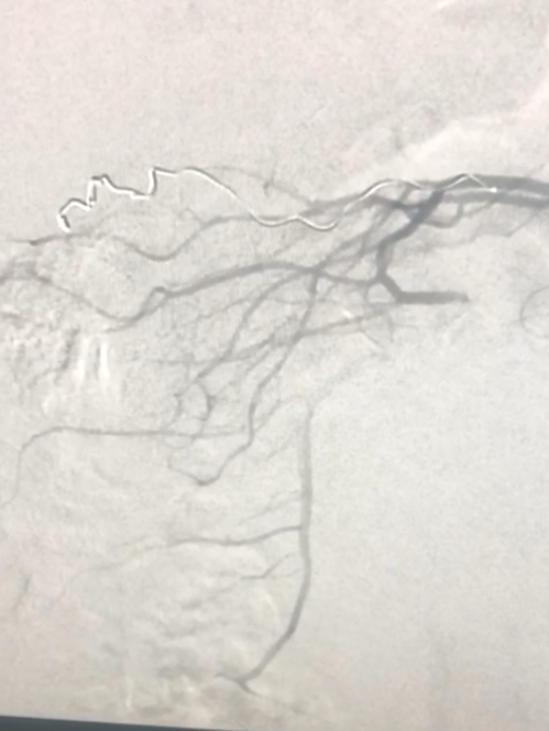
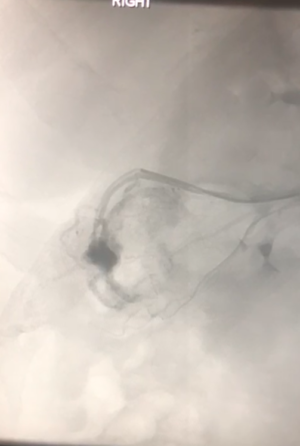
1-Blood vessel disease:
Arteries:
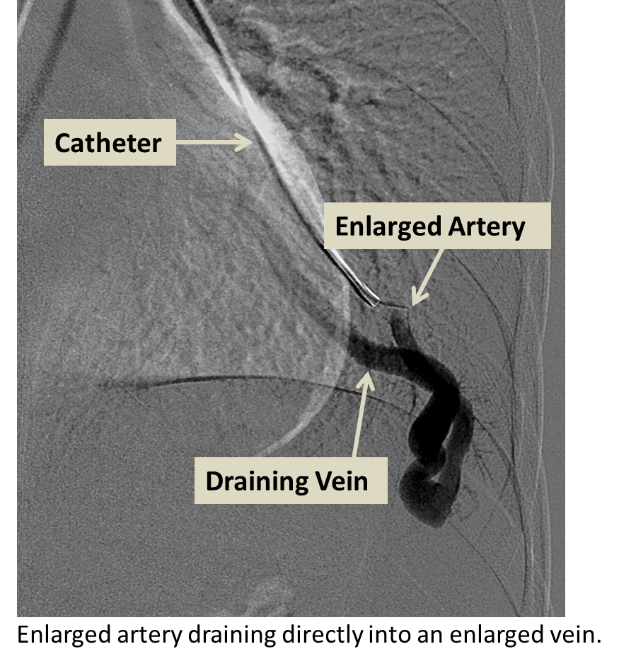
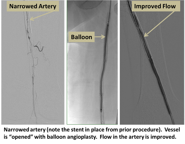
Narrowing of arteries leading to restricted blood flow (peripheral vascular disease): lnterventional radiologists treatthis by using balloons to stretch the vessel (balloon angioplasty, PTA) and sometimes metal springs called stents tohold them open. Sometimes arteries or bypass grafts block suddenly with a rapid loss of blood supply to the limb. Unless the bloodsupply is restored this can lead to amputation. lnterventional radiologists can help by infusion of clot busting drugsdirectly into the artery via small catheters thus saving many limbs. Expanded arteries (aneurysms) at risk of rupture and bleeding: IRs treat these by relining the vessel with a tube called a stent graft = Bleeding (haemorrhage). This is the most common vascular emergency treated by IR. Haemorrhage can come from almost anywhere e.g. from the gut, secondary to major injury or following birth. Bleeding can often permanently be stoppedby blocking the vessel (embolization), relining the vessel with a stent graft or by blowing up a balloon in the vessel to stop the bleeding until emergency surgery can be performed. lnterventional radiology is also used to prevent bleedingduring some sorts of surgery e.g. during caesarean section in patients with a high risk of bleeding from an abnormalplacenta (post partum haemorrhage).
Arteries:
Narrowing of arteries leading to restricted blood flow (peripheral vascular disease): lnterventional radiologists treatthis by using balloons to stretch the vessel (balloon angioplasty, PTA) and sometimes metal springs called stents tohold them open. Sometimes arteries or bypass grafts block suddenly with a rapid loss of blood supply to the limb. Unless the bloodsupply is restored this can lead to amputation. lnterventional radiologists can help by infusion of clot busting drugsdirectly into the artery via small catheters thus saving many limbs. Expanded arteries (aneurysms) at risk of rupture and bleeding: IRs treat these by relining the vessel with a tube called a stent graft = Bleeding (haemorrhage). This is the most common vascular emergency treated by IR. Haemorrhage can come from almost anywhere e.g. from the gut, secondary to major injury or following birth. Bleeding can often permanently be stoppedby blocking the vessel (embolization), relining the vessel with a stent graft or by blowing up a balloon in the vessel to stop the bleeding until emergency surgery can be performed. lnterventional radiology is also used to prevent bleedingduring some sorts of surgery e.g. during caesarean section in patients with a high risk of bleeding from an abnormalplacenta (post partum haemorrhage).

2-Non vascular intervention:
This is sometimes referred to as interventional oncology but the treatments are also effective in benign conditions. IR therapies are used for the following: Tumour therapies:
This is sometimes referred to as interventional oncology but the treatments are also effective in benign conditions. IR therapies are used for the following: Tumour therapies:
•to treat the tumour I cancer (tumour ablation, embolization)
•to relieve the effects of the cancer on other systems e.g. blockage of the gullet (oesophagus), bowel, kidney
(nephrostomy) or liver (biliary drainage)
•To drain collections of fluid or pus in the chest or abdomen
•To place feeding tubes (gastrostomy, jejunostomy)
•To treat collapsed spinal bones (vertebroplasty)
these treatments are intended to shrink or destroy
tumours at their primary site or which have spread to other areas (metastases).
This is an area of increasing interest and leading to improved survival with reduced morbidity.
Liver, kidney and other tumours (e.g. bone, lung): these can be treated by destructive therapies(ablation) usually involving heat (radiofrequency, laser,
microwave, ultrasound) or cold damage (cryotherapy). The treatment is performed and monitored usingimaging (ultrasound, computed tomography or magnetic resonance imaging).
Uterine fibroids:
heavy menstrual bleeding and pain can be caused bybenign tumours called fibroids.
These can be treated by blocking blood vessels (uterine fibroid embolization, UFE) which leads to shrinkage.
Embolization is sometimes combined with drug therapy (chemoembolization) or radiotherapy(radioembolization) which targets the effect to the tumour and limits some of the side effects of cancer therapy.
3. Some of our procedures